Advertisement

We need your help now

Support from readers like you keeps The Journal open.

You are visiting us because we have something you value. Independent, unbiased news that tells the truth. Advertising revenue goes some way to support our mission, but this year it has not been enough.

If you've seen value in our reporting, please contribute what you can, so we can continue to produce accurate and meaningful journalism. For everyone who needs it.
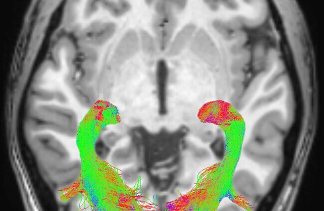
MRI of the brain Radiological Society of North America

Scientists find visual symptoms that could help early detection of Parkinson's

Non-motor Parkinson’s symptoms may precede the appearance of motor signs by more than a decade.

CHANGES IN THE how a newly-diagnosed Parkinson’s patient sees things could provide important biomarkers for early detection of the disease.

Researchers have found “significant abnormalities” within the visual system brain structures of Parkinson’s patients which they believe can precede motor signs by a decade.

Parkinson’s disease is a progressive disease of the nervous system caused by neuronal loss in the brain structure.

The disease is characterised by tremors, muscular rigidity and slow, imprecise movements.

Non-motor symptoms experienced by people with Parkinson’s include visual alterations such as an inability to perceive colours, a change in visual acuity, and a decrease in blinking with can lead to dry eyes, according to researchers.

“Although Parkinson’s disease is primarily considered a motor disorder, several studies have shown non-motor symptoms are common across all stages of the disease,” lead researcher Dr Alessandro Arrigo said.

These symptoms are often undiagnosed because patients are unaware of the link to a disease and, as a result, they may be under-treated.

“These non-motor Parkinson’s symptoms may precede the appearance of motor signs by more than a decade,” he said.

The researchers studied 20 newly diagnosed and not yet treated patients – 11 men and nine women – with Parkinson’s, and 20 age and gender-matched healthy patients.

MRI scans were performed on both groups.

Researchers used an MRI technique called diffusion weighted imaging to assess white matter changes to investigation changes of the brain’s grey and white matter.

The results showed significant abnormalities in the visual system brain structures of the Parkinson’s patient group.

These abnormalities included alterations of optic radiations, a reduction of white matter concentration and a reduction of optic chiasm volume.

“Just as the eye is a window into the body, the visual system is a window into brain disorders,” Arrigo said.

Visual processing metrics may prove helpful in differentiating Parkinson’s disorders, following disease progression, and monitoring patient response to drug treatment.

Arrigo added that future studies are needed to better understand the timing of degeneration in a visual sense, as well as the specific changes that occur.

Read: In 2014, a case similar to Charlie Gard’s came before the Irish High Court

More: Tobacco giant Philip Morris ordered to pay Australian government millions

Readers like you are keeping these stories free for everyone...
A mix of advertising and supporting contributions helps keep paywalls away from valuable information like this article. Over 5,000 readers like you have already stepped up and support us with a monthly payment or a once-off donation.

Close
5 Comments
    Submit a report
    Please help us understand how this comment violates our community guidelines.
    Thank you for the feedback
    Your feedback has been sent to our team for review.
    JournalTv
    News in 60 seconds