Advertisement

We need your help now

Support from readers like you keeps The Journal open.

You are visiting us because we have something you value. Independent, unbiased news that tells the truth. Advertising revenue goes some way to support our mission, but this year it has not been enough.

If you've seen value in our reporting, please contribute what you can, so we can continue to produce accurate and meaningful journalism. For everyone who needs it.
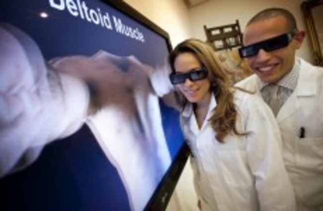
Students experience the new 3D anatomical aid through special goggles at the RHA yesterday Andres Poveda via apphoto.ie

Ireland develops world's first 3D surface anatomy guide

A team of artists, anatomists and engineers have proven that 3D is not just useful for blockbuster movies – it can stand in for “live models” too.

WHAT DO YOU get when you combine artists, anatomists and engineers? The world’s first 3D surface anatomy guide, of course.

A collaboration between these three groups over the past two years in Ireland has let to an online 3D guide “which shows the motions of muscles and the sites of structures from the surface inwards”. This is the first such guide to be created in the world and will be used by medical students, artists and surgical students as a training aid.

The project, which uses 3D technology to bring the anatomical structures to virtual life, was funded by the Science Foundation Ireland and the Royal College of Surgeons Ireland (RCSI), and shown off for the first time at the Royal Hibernian Academy in Dublin yesterday. The computer-based training aid will become part of the curriculum for med students in Dublin, Bahrain and Kuala Lumpur immediately. It will be used to teach anatomy to artists in the RHA school from next year.

According to its creators, the 3D model will help lecturers get round the lack of “willing live models” to show off their bodies. Professor Clive Lee, head of the Department of Anatomy at the RCSI, said that artists and models do not always have access to live models whenever they want to examine surface anatomical structure. He said:

Medical students are reluctant models, so getting one to take off his shirt to demonstrate the surface anatomy of the heart valves can be a challenge. Yet surface anatomy is the basis of clinical examination and students must learn where to listen to the heart, the markings of the liver, the sites of incisions and the movements of joints – in short the site of everything from the outside in.

Artists also do not always have access to a live model either, and for them a knowledge of the underlying skeletal and muscular structure is fundamental to drawing and painting the figure.This guide allows us to teach these vital lessons in a richer and more engaging manner and on a more frequent basis.

The team of researchers behind the project include Professor Lee, Dr Valerie Morris of the RCSI, engineers David Corrigan and Anil Kokaram, and artists Mick O’Dea and Una Sealy. It makes use of stereoscopic-3D, a technology that creates the illusion of depth in a two-dimensional image. And yes, to view the new anatomical aid, users will have to don those special 3D goggles (see main pic, above).

This is the tool online – wearing the goggles adds depth of field to viewing the anatomical structure:

Readers like you are keeping these stories free for everyone...
A mix of advertising and supporting contributions helps keep paywalls away from valuable information like this article. Over 5,000 readers like you have already stepped up and support us with a monthly payment or a once-off donation.

Close
5 Comments
    Submit a report
    Please help us understand how this comment violates our community guidelines.
    Thank you for the feedback
    Your feedback has been sent to our team for review.
    JournalTv
    News in 60 seconds