Advertisement

We need your help now

Support from readers like you keeps The Journal open.

You are visiting us because we have something you value. Independent, unbiased news that tells the truth. Advertising revenue goes some way to support our mission, but this year it has not been enough.

If you've seen value in our reporting, please contribute what you can, so we can continue to produce accurate and meaningful journalism. For everyone who needs it.
Pics: 11 curious close-ups of scientific images

The Health Research Board wants to “showcase the magic of science” with amazing images. Job done.

THIS BEING THE month that the Euroscience Open Forum hits Dublin (see #esof2012 hashtag on Twitter too), now’s as good a time as any to find the “magic” in science.

To that end, the national Health Research Board has been doing its bit, compiling amazing MRI and other close-up images on its Flickr stream

If you’ve been to Dublin recently, you might have seen some of the images blown up on the front of some city buildings on Baggot Street Bridge, Pearse Street and on Liberty Hall.

Pics: 11 curious close-ups of scientific images
1 / 8
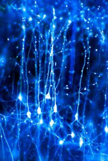
  • Neurons in the brain

    This image is a colour-enhanced light microscopy image of pyramidal neurons forming a network in the brain. Pyramidal neurons are so-called because they have a pyramid-shaped cell body (soma) and also have very long branches (dendrites) and have been found in birds, fish, reptiles, and all mammals. They are found in the front forebrain of mammal’s brains and are thought to be involved in a person's ability to process thoughts. If these neurons no longer function properly, this is likely to lead to diseases such as Alzheimer’s disease or schizophrenia. Credit: Jonathan Clarke © Wellcome Images. Creative Commons
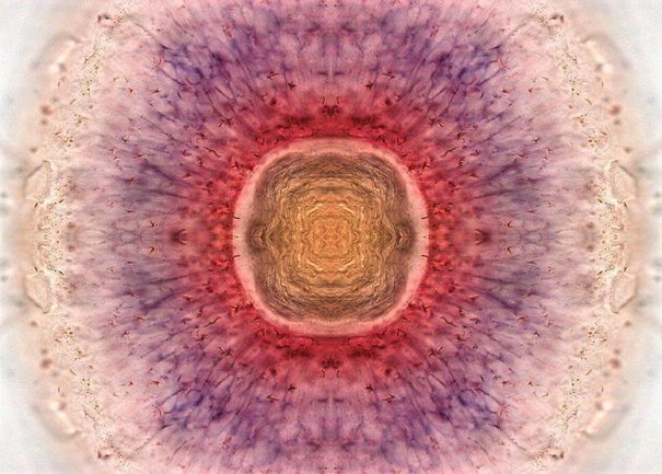
  • A zebrafish retina

    This image shows the retina from the eye of a three-day-old zebrafish - small tropical freshwater fish that are widely used in scientific research. It was created using a staining technique called double in situ hybridisation where different structures of the eye can be identified by staining for genes known to be found in specific tissues. The central yellow region of the image is the lens of the zebra fish eye. Stem cells are stained in purple and red, with the purple cells beginning to differentiate into the functional neurons which are necessary for sending visual signals to the brain. Credit: Kara Cerveny & Steve Wilson Copyright © Wellcome Images. Creative Commons.
  • Ruptured blood vessel

    This is a colour-enhanced scanning electron micrograph of red blood cells leaking out of a ruptured blood vessel. The leaking of blood is due to a mutation in a particular gene (the ephrin-B2 gene) that causes the blood vessels to be more fragile than normal, leading to an increased rate of bleeding. The fragility is due to the inadequate coverage of the blood vessel by smooth muscle cells. This kind of leaky blood vessel is frequently found in tumours and in certain other human diseases. Image Credit: Anne Weston. © Wellcome Images. Creative Commons.
  • The Hive

    This image shows the porous surface of a biodegradable polymer (PLGA) which is used in a variety of medical devices such as implants, sutures, and artificial limbs. These porous biomaterials could have applications in areas such as drug delivery and wound care. Image Credit: David McGovern, CRANN, Trinity College Dublin Copyright © Science Foundation Ireland
  • MRSA

    Though this may appear to be a bunch of grapes, these are actually clusters of methicillin-resistant Staphylococcus aureus (MRSA) bacteria, visualised using colour-enhanced scanning electron micrograph techniques. Known as the “hospital superbug”, MRSA is resistant to some of the most powerful antibiotics available and thrives in places like hospitals where people often have open wounds and weakened immune systems which put them at greater risk than normal. Image Credit: Annie Cavanagh. © Wellcome Images. Creative Commons.
  • Celtic Knot

    This is an image of the 3D polymeric biomaterial known as a “Celtic Knot”. Research using biomaterials such as this one involves developing implantable materials to restore and regenerate cells and tissue that result from disease or injury. Hard-to-heal chronic wounds affect millions of patients each year, such as those suffering from Epidermolysis Bullosa (EB). The Celtic Knot polymer, with its high degree of branching (in grey) and multiple functionalities (green and blue points) could be used to deliver drugs within a new type of dressing to speed up wound healing. Image Credit: Wenxin Wang Copyright © Science Foundation Ireland
  • Enhanced MRSI scan of the head

    This image is a digitally enhanced Magnetic Resonance Imaging (MRI) scan of a human head showing the brain and spinal cord in orange/yellow and the other tissues in blue and pink. MRI scans can record brain activity as it unfolds over time. It is founded on the fact that highly active neurons in the brain consume more oxygen than those that are less active. The oxygen rich blood that is sent to the more active regions of the brain has slightly different magnetic properties to blood that has given up its oxygen to the surrounding tissue. This difference gives rise to the blood oxygenation level–dependent (BOLD) signal and can be recorded with magnetic resonance imaging. Image Credit: Mark Lythgoe , Chloe Hutton. © Wellcome Images. Creative Commons.
  • Invisible Whirling Whip

    This is a dinoflagellate (from the Greek dinos "whirling" and Latin flagellum "whip") captured in Dublin Bay, and seen using colour-enhanced scanning electron microscope techniques. Dinoflagellates are a prominent member of the plankton family which are found in both marine and freshwater ecosystems. The most dramatic effect of dinoflagellates on their environment occurs in coastal waters where they can grow so much as to turn the water red (Red tide) or luminescent (blue-green) at night. However, they are also extremely poisonous and their toxins can be quickly carried up the food chain and passed onto humans if they eat contaminated fish and shellfish, which may result in gastrointestinal illness, permanent neurological damage, or even death. This image is part of a series of exhibitions, movies and talks about the Tara Oceans Expedition in Dun-Laoghaire and at ESOF 2012 supported by the French Embassy in Ireland. Copyright © Aine McKeon, Anika Mostaert, Emmanuel G. Reynaud - Tara Oceans - UCD, 2009

Readers like you are keeping these stories free for everyone...
A mix of advertising and supporting contributions helps keep paywalls away from valuable information like this article. Over 5,000 readers like you have already stepped up and support us with a monthly payment or a once-off donation.

Close
6 Comments
    Submit a report
    Please help us understand how this comment violates our community guidelines.
    Thank you for the feedback
    Your feedback has been sent to our team for review.
    JournalTv
    News in 60 seconds